
 This article considers how clinical photographs benefit both the clinician and the patient, and looks at the key features we want to capture when taking these pictures.
Why we take clinical photographs in dentistry
The main purpose of taking clinical photographs in dentistry is to make an accurate record of the clinical findings in a patient’s mouth. The photographs are useful to both the clinician and the patient, and can be used in a number of ways:
- They help the clinician plan treatment (alongside other forms of record-taking, such as radiographs and charting).
- They provide a baseline reference point for the patient’s mouth at the start of treatment. The clinician can then refer to these initial pictures when monitoring the treatment’s progress – for example, during teeth-whitening, orthodontics, restorative work and implantology.
- They can be used as motivational tool for patients during treatment. Pictures demonstrating progress are a great source of encouragement.
- Taking clinical photographs at different stages of treatment help us as dental professionals to communicate with patients more effectively – visual aids are very useful when discussing findings and progress.
- The photographs can be used as a medico-legal record. It is recommended that clinicians keep photographic records of any procedures that could lead to litigation.
- The use of photographs can help communication between the clinician and other health care professionals – for example, if a patient is referred to another specialist for treatment.
- They enhance staff training. Photographs are a great visual aid in educating and training staff in dentistry.
- Clinical photographs are good for practice marketing.
Consistently good clinical photographs
In the practice where I work, we have procedures in place to ensure we take consistently good clinical photographs. We:
- Use a high-quality camera and flash, keeping up to date with new technology.
- Have regular staff training.
- Conduct routine audits to monitor picture quality.
- Communicate effectively with our patient.
- Have the help of an assistant for retraction.
- Use standardisation for comparison at different times; for example, by using the same background in pictures and by having a consistent camera magnification and a consistent distance between the patient and the photographer.
Taking clinical photographs
When taking clinical photographs, we need:
- Patient consent.
- A camera.
- The patient.
- Retractors and mirrors.
- An assistant for help with retraction.
Before we start taking the photographs, we need to prepare the patient. Coats, scarves, hats and glasses should be removed. The patient’s hair should be tucked behind their ears if it is likely to obstruct areas of interest. The patient’s head should be positioned correctly.
In the dental surgery, the different types of clinical photographs taken are:
Extra-oral (outside the patient’s mouth)
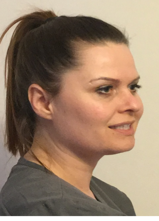
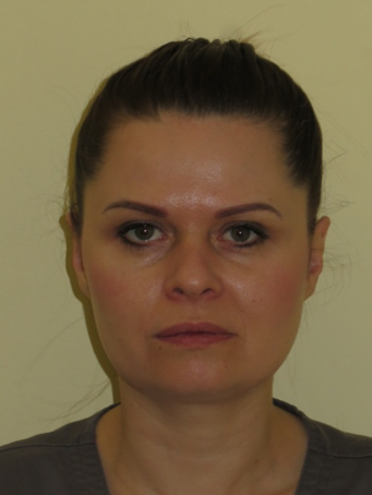 1) Face frontal
This is a picture of the patient’s face looking straight at the camera. The patient should have a neutral expression and their lips at rest.
When taking this picture, we aim to capture the patient’s face from the top of the crown to the collar bone.
The photograph should be symmetrical.
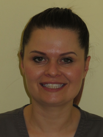 2) Face smiling
This is a picture of the patient’s face looking straight at the camera. The patient’s position is the same as for the face frontal photograph – the only difference is that the patient will be asked to smile.
This photograph will help to record soft tissue and facial aesthetics when the patient is smiling.
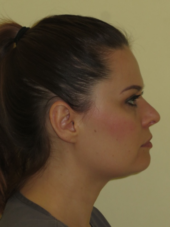 3) Profile picture
For this photograph, the patient will be asked to turn to the left and face forward so that their profile can be captured.
The aim for this photograph is to capture the patient’s face from the top of their crown to the collar bone.
The Frankfort plane should be horizontal to ensure the patient is looking straight ahead.
 4) 45-degree angle profile
After having the profile picture taken, the patient is asked to turn their head slightly to the right at a 45-degree angle. In this picture, the patient’s teeth should be visible. The aim is to capture visible information about the patient’s smile.
Intra-oral (inside the patient’s mouth)
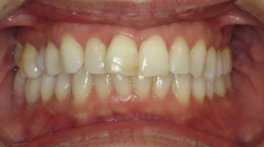 1) Frontal – in occlusion
For this photograph, the patient is asked to take a seat in the dental chair. The chair’s height is adjusted so that it is level with the clinician’s elbows. An assistant stands behind the patient to retract the patient’s lips sideways. The patient is asked to keep their teeth biting together, and a photograph is then taken at a 90-degree angle to the facial midline.
In this photograph, the aim is for the central incisors to be positioned centrally with the buccal corridors visible.
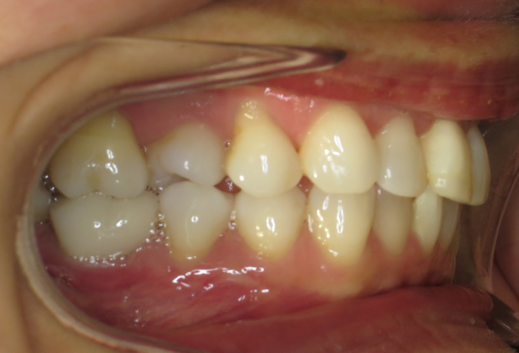 2) Right buccal – in occlusion
Retractors are used to help retract the patient’s lips and cheeks. The patient is asked to turn their head all the way to the left so that their ear is touching the headrest. The assistant who is helping with retraction then pulls on the right retractor so that the right-hand-side teeth are exposed in occlusion.
In this photograph, the aim is to capture the mesial aspect of the left central incisor to the first molar and further back if possible whilst teeth are in occlusion.
This photograph should be taken from a 90-degree angle.
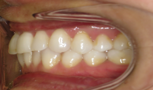 3) Left buccal – in occlusion
Retractors are used to help retract the patient’s lips and cheeks. The patient is asked to turn their head all the way to the right so that their ear is touching the headrest. The assistant who is helping with retraction then pulls on the left retractor so that the left-hand-side teeth are exposed in occlusion.
In this photograph, the aim is to capture the mesial aspect of the right central incisor to the first molar and further back if possible whilst teeth are in occlusion.
This photograph should be taken from a 90-degree angle.
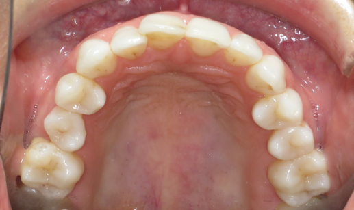
 4) Occlusal – upper
For this picture, we use a mirror, which is warmed slightly with warm water to help prevent fogging.
Two retractors are used to hold the patient’s top lip out of the way. The patient can help by holding the retractors whilst the assistant positions the mirror. The mirror is then moved to the back of the mouth and angled so that the upper occlusal arch can be captured.
The photograph should show the buccal and palatal surfaces of the incisors.
The image should start in front of the incisors, extending back to a minimum of the distal surface of the first molar, but ideally capturing all erupted teeth in the arch.
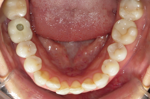 5) Occlusal – lower
Two retractors are used to hold the patient’s lower lip out of the way. The warmed mirror (see above) is then moved to the back of the mouth and angled so that the lower occlusal arch can be captured. To help ensure all required elements are captured, we ask the patient to relax their tongue. If they are unable to do this, we ask them to try to curl it to the back of the mouth so that the mirror can rest in front.
The photograph should show the buccal and lingual surfaces of the incisors.
As with the upper occlusal, the image should start in front of the incisors, extending back to a minimum of the distal surface of the first molar but ideally capturing all erupted teeth in the arch.
The upper and lower photographs should be symmetrical views of the occlusal surfaces of the teeth.
When taking occlusal views, it is important to warn the patient that the mirror is made from glass and that they should be careful not to bite down on it.
Conclusion
In the orthodontic practice where I work, we take clinical photographs daily and use them to plan treatment and monitor patient progress.
Taking clinical photographs benefits the clinician and patient, but also benefits us as nurses. Taking photographs gives us the opportunity to learn new skills and allows us to be more hands-on with patients, which is rewarding.
Having a variety of skills makes you a valued member of the dental team and gives a great sense of job satisfaction.
Speaking from personal experience, I have enjoyed learning how to take clinical photographs. I found it a lot harder than I expected, but like many things, practice does make perfect.
As clinical photography plays such a key role in all aspects of dentistry, I am pleased that I am now competent at taking clinical photographs, as this is a skill that I will use throughout my career in dentistry.
 Written by Emma Edwards RDN
NEBDN National Certificate in Dental Nursing, NEBDN Dental Sedation Nursing, NEBDN Oral Health Education , NEBDN Dental Radiography, Kings Certificate in Implant Nursing, Level 5 Diploma in Leadership and Management, Level 3 Award in Education and Training.